Brain MRI Classification in collaboration with Kantonsspital Winterthur

Brain MRI Classification in collaboration with Kantonsspital Winterthur
Project by: Cornelia Schmitz, Norbert Bräker
 |
 |
| Cornelia Schmitz | Norbert Bräker |
Introduction
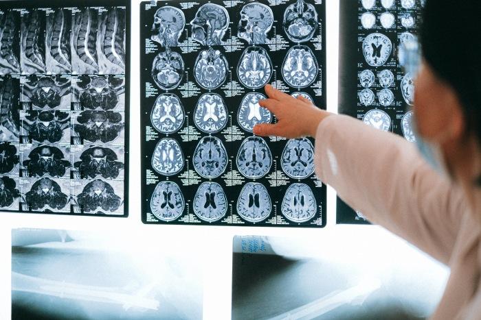
This objective of the project was to automate the detection and classification of brain tumors using Magnetic Resonance Imaging (MRI) with the aid of advances in Artificial Intelligence, particularly Neural Networks. The term "brain tumor" covers both the various types that develop directly in the brain and brain metastases, i.e., metastases of a tumor that originally grew outside the brain. Detecting tumors of the brain and making an assessment of the type is nowadays done by radiologists who can usually do this reliably with the help of Magnetic Resonance Imaging (MRI). Automating this process, however, has the advantage of being used as a second opinion, further reducing the risk of overlooking a small tumor or misclassifying one. Likewise, automatic detection can assist the radiologist by accurately documenting the position of multiple metastases, for example, when multiple occurrences are present (Source: SNFS).
Project details
Cornelia Schmitz and Norbert Bräker built a classification network to group images based on two properties: the perspective and the MR sequence used to generate each image. The task our students were given is based on previous work from Paul Windisch, a Physician at Kantonsspital Winterthur and former Data Science student at SIT Academy. His idea was to have a pipeline of networks which are used for brain tumor detection. As inputs, Cornelia and Norbert were given MR images and as an output, they were tasked to predict the sequence and perspective of the image.
At the beginning of this pipeline of networks, the task was to classify new features that will then be transferred to the next network. The two main features our students worked with were perspective (side, front, top) and sequence. The MR sequence determines which tissues appear lighter or darker (evident on image below). The images provided were first grouped and then directed into specialized networks with the goal of improving the quality of tumor classification and segmentation.

The first approach that was used is called Transfer Learning. The idea was to use a Neural Network that has been previously trained on millions of images. The advantage of this approach is that you need much fewer images than if you had to train the network from scratch. A total of 409 images were used, which first had to be manually selected and labeled. These were then fed into the previously trained network, to which additional layers were added, to allow the network to learn specifically about MR images. As a result, our students were given the perspective and sequence of the images.
Results of transfer learning:

Now that the network is fully trained with the available images, time can be saved since they do not need to be selected manually.
At the beginning of this pipeline of networks, the task was to classify new features that will then be transferred to the next network. The two main features our students worked with were perspective (side, front, top) and sequence. The MR sequence determines which tissues appear lighter or darker (evident on image below). The images provided were first grouped and then directed into specialized networks with the goal of improving the quality of tumor classification and segmentation.

The first approach that was used is called Transfer Learning. The idea was to use a Neural Network that has been previously trained on millions of images. The advantage of this approach is that you need much fewer images than if you had to train the network from scratch. A total of 409 images were used, which first had to be manually selected and labeled. These were then fed into the previously trained network, to which additional layers were added, to allow the network to learn specifically about MR images. As a result, our students were given the perspective and sequence of the images.
Results of transfer learning:

Now that the network is fully trained with the available images, time can be saved since they do not need to be selected manually.
Conclusion
In conclusion, an approach has been found that can correctly classify, segment, and group the images. During this project, some new interesting architectures were found that our students will continue to explore to potentially improve the performance of the models even further. The classification accuracy of the Transfer Learning model was between ~94% - 98% for perspective and sequence. The project as a whole showed that the performance of an algorithm for brain tumor classification is satisfactory, but not yet sufficient for routine clinical application. Further research will now investigate whether better results can be achieved with more complex Neural Networks on the data sets used. The work done in this project will help Paul Windisch in his own research on automatic brain tumor detection, a project he started with Pascal Weber during his studies at SIT Academy and now funded by SNSF (Swiss National Science Foundation) and Innosuisse (Swiss Agency for Innovation Promotion).
Find more information about the project details on Cureus.
Student
Cornelia Schmitz says:
Joining SIT Academy helped me develop my skills and gave me the confidence to apply for interesting roles in Data Science. Through the experiences I gained in the bootcamp, I was able to land a job at the tech startup Eyeware.
Hello world